File:Pisiform fracture (Radiopaedia 52465).jpeg
Jump to navigation
Jump to search

Size of this preview: 459 × 600 pixels. Other resolutions: 184 × 240 pixels | 466 × 609 pixels.
Original file (466 × 609 pixels, file size: 87 KB, MIME type: image/jpeg)
Summary:
- Radiopaedia case ID: 52465
- Image ID: 29606778
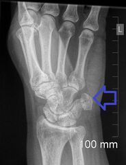
- Study findings: AP and lateral view demonstrated no acute abnormality. Additional lateral 30 degree angled view was requested given patient's symptoms which demonstrated fracture of the pisiform bone. No other fracture was identified.
- Modality: X-ray
- System: Musculoskeletal
- Findings: AP and lateral view demonstrated no acute abnormality. Additional lateral 30 degree angled view was requested given patient's symptoms which demonstrated fracture of the pisiform bone. No other fracture was identified.
- Published: 10th Apr 2017
- Source: https://radiopaedia.org/cases/pisiform-fracture-4
- Author: Aneta Kecler-Pietrzyk
- Permission: http://creativecommons.org/licenses/by-nc-sa/3.0/
Licensing:
Attribution-NonCommercial-ShareAlike 3.0 Unported (CC BY-NC-SA 3.0)
File history
Click on a date/time to view the file as it appeared at that time.
| Date/Time | Thumbnail | Dimensions | User | Comment | |
|---|---|---|---|---|---|
| current | 06:09, 24 September 2022 | | 466 × 609 (87 KB) | Doc James (talk | contribs) | Added arrow |
| 12:27, 26 March 2021 | 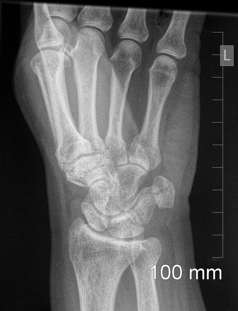 | 466 × 609 (171 KB) | Fæ (talk | contribs) | Radiopaedia project rID:52465 (batch #29414) |
You cannot overwrite this file.
File usage
There are no pages that use this file.
.jpeg&oldid=8852648)