File:Pntd.0003908.g002.png
Jump to navigation
Jump to search
No higher resolution available.
Pntd.0003908.g002.png (600 × 428 pixels, file size: 232 KB, MIME type: image/png)
Summary
| Description |
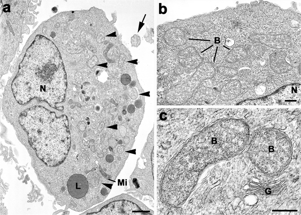
English: Electron microscopic appearance of N. sennetsu (ATCC VR-367, Miyayama strain) in DH82 canine monocyte cultures. a)
Low power micrograph of an infected cell in which a number of bacteria can be identified in the cytoplasm (arrowheads) in addition to the nucleus (N), mitochondria (Mi) and lipid droplet (L). Note the single extracellular bacterium (arrow). Bar represents 1μm. b) Enlargement of part of the cytoplasm showing a number of gram negative bacteria (B). N–nucleus. Bar represents 200nm. c) Detail of N. sennetsu (arrow) showing the gram negative bacteria limited by two unit membranes located within a membrane bound vacuole. G—Golgi stack. Bar represents 200nm. |
| Date | |
| Source | https://www.ncbi.nlm.nih.gov/pmc/articles/PMC4497638/ |
| Author | Sabine Dittrich, Weerawat Phuklia, Gareth D. H. Turner, Sayaphet Rattanavong, Vilada Chansamouth, Stephen J. Dumler, David J. P. Ferguson, Daniel H. Paris, Paul N. Newton |
Licensing
English: This file is licensed CC BY-NC 4.0
This file was uploaded with UploadWizard.
File history
Click on a date/time to view the file as it appeared at that time.
| Date/Time | Thumbnail | Dimensions | User | Comment | |
|---|---|---|---|---|---|
| current | 20:14, 11 May 2023 | | 600 × 428 (232 KB) | Ozzie10aaaa (talk | contribs) | Uploaded a work by Sabine Dittrich, Weerawat Phuklia, Gareth D. H. Turner, Sayaphet Rattanavong, Vilada Chansamouth, Stephen J. Dumler, David J. P. Ferguson, Daniel H. Paris, Paul N. Newton from https://www.ncbi.nlm.nih.gov/pmc/articles/PMC4497638/ with UploadWizard |
You cannot overwrite this file.
File usage
There are no pages that use this file.