Category:UndergradImaging
Jump to navigation
Jump to search
Photos from : Undergraduate Diagnostic Imaging Fundamentals
Subcategories
This category has the following 114 subcategories, out of 114 total.
A
- Abdomen (UndergradImaging)
- Acromioclavicular Joint Separation (UndergradImaging)
- Angiography (UndergradImaging)
- Ankle Trauma, Fractures (UndergradImaging)
- Aortic Dissection and Aneurysm (UndergradImaging)
- Appendicitis (UndergradImaging)
- Approach to Bone and Joint x-rays (UndergradImaging)
- Approach to the Abdominal x-ray AXR (UndergradImaging)
- Approach to the Chest x-ray CXR (UndergradImaging)
- Appropriateness of Imaging Guidelines (UndergradImaging)
- Atelectasis (UndergradImaging)
B
C
- Cardiovascular – References (UndergradImaging)
- Chest (UndergradImaging)
- Chest – References (UndergradImaging)
- Cholecystitis (UndergradImaging)
- Choosing Wisely – Canada (UndergradImaging)
- Classification of Radiation Damage (UndergradImaging)
- Clavicle Fracture (UndergradImaging)
- Computed Tomography (UndergradImaging)
- Congestive Heart Failure (UndergradImaging)
- Contrast Media in Radiology (UndergradImaging)
E
F
G
H
I
- Ileus (UndergradImaging)
- Image Gently (UndergradImaging)
- Inferior Vena Cava Filter (UndergradImaging)
- Interventional/Vascular – References (UndergradImaging)
- Intestinal Obstruction (UndergradImaging)
- Intestinal Perforation- Pneumoperitoneum (UndergradImaging)
- Intracranial Hemorrhage – Traumatic (UndergradImaging)
- Introduction (UndergradImaging)
- Introduction to Breast Imaging (UndergradImaging)
- Introduction – References (UndergradImaging)
- Ionizing Radiation: Basic Concepts (UndergradImaging)
- Ionizing Radiation: Fetus and Neonate (UndergradImaging)
- Ischemic Stroke (UndergradImaging)
L
M
N
P
- Palpable Breast Mass (UndergradImaging)
- Pediatric (UndergradImaging)
- Pediatric – References (UndergradImaging)
- Pelvic Fracture (UndergradImaging)
- Pelvis (UndergradImaging)
- Percutaneous Biopsy (UndergradImaging)
- Percutaneous Fluid Drainage (UndergradImaging)
- Permissions (UndergradImaging)
- Placenta Previa (UndergradImaging)
- Pleural Effusion (UndergradImaging)
- Pneumonia (UndergradImaging)
- Pneumothorax (UndergradImaging)
- Principles of Imaging Techniques – References (UndergradImaging)
- Principles of Radiation Biology and Radiation Protection – References (UndergradImaging)
- Pulmonary Thromboembolism (UndergradImaging)
- Pyloric Stenosis (UndergradImaging)
R
- Radiation in Medical Imaging: The x-ray Tube (UndergradImaging)
- Radiation Interaction with Biological Matter (UndergradImaging)
- Radiation Protection for Healthcare Workers (UndergradImaging)
- Radiation Protection for Patients (UndergradImaging)
- Renal Tumour (UndergradImaging)
- Retropharyngeal Abscess – Child (UndergradImaging)
- Rotator Cuff (UndergradImaging)
S
T
U
Media in category "UndergradImaging"
The following 200 files are in this category, out of 355 total.
(previous page) (next page)-
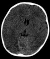
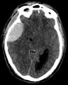 6.2A Axial CT of Head displaying epidural hematoma.jpg 866 × 1,070; 125 KB
-
 Abdomen Plain x-ray, Labelled.png 424 × 455; 229 KB
-
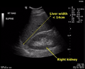
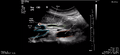 Abdomen US – Liver and Abdominal Midline.png 1,017 × 462; 194 KB
-
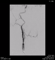 Abnormal CTA 1.png 675 × 742; 255 KB
-
 Abnormal CTA 2.png 675 × 742; 291 KB
-
 Abnormal CTA 3.png 675 × 742; 304 KB
-
 Abnormal CTA 4.png 675 × 742; 344 KB
-
 Annotated Normal Pediatric Lateral Chest x-ray.png 675 × 742; 557 KB
-
 Annotated Normal Pediatric PA Chest x-ray.png 675 × 742; 588 KB
-
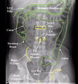
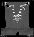
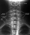 AP Adult C-spine, Labelled.jpeg 432 × 501; 52 KB
-
 Axial CT 1.png 675 × 791; 144 KB
-
 Axial CT 2.png 675 × 742; 150 KB
-
 Coronal CT 1.png 675 × 742; 328 KB
-
 Coronal CT 2.png 675 × 742; 299 KB
-
 Fetal Head with measurements.png 683 × 742; 241 KB
-
 Fetal Kidneys with measurements.png 683 × 742; 234 KB
-
 Fetal Legs with femur measurements.png 683 × 742; 270 KB
-
 Fetal Orbits with measurements.png 683 × 742; 270 KB
-
 Fetal spine with measurements.png 683 × 742; 265 KB
-
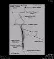
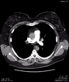 Fig 17.40A Normal CTA of Chest.png 675 × 791; 300 KB
-
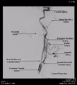
 Fig 17.40B Normal CTA of Chest.png 675 × 742; 303 KB
-
 Figure 1.1 ODIN Search Page.jpg 2,336 × 1,322; 162 KB
-
 Figure 1.10 ODIN Download Image Instructions Pane.jpg 2,247 × 1,761; 290 KB
-
 Figure 1.11 ODIN Image Stack.jpg 2,206 × 1,314; 266 KB
-
 Figure 1.12 ODIN Looping Speed Tool.jpg 366 × 96; 7 KB
-
 Figure 1.15 ODIN Zoom Tool.jpg 798 × 150; 12 KB
-
 Figure 1.16 ODIN Zoomed In Image View.jpg 2,186 × 1,704; 321 KB
-
 Figure 1.17 ODIN Reset Image Tool.jpg 633 × 132; 8 KB
-
 Figure 1.19a ODIN Image Layout Tool (multiple).jpg 2,254 × 1,752; 403 KB
-
 Figure 1.19b ODIN Image Layout Tool (single).jpg 2,260 × 1,776; 343 KB
-
 Figure 1.2 ODIN Default Image View.jpg 2,234 × 1,772; 475 KB
-
 Figure 1.20 ODIN Text Overlay Tool.jpg 2,283 × 1,812; 348 KB
-
 Figure 1.21a ODIN View Metadata Tool (button).jpg 627 × 138; 17 KB
-
 Figure 1.21b ODIN View Metadata Tool (dialog box).jpg 2,247 × 1,797; 322 KB
-
 Figure 1.22a ODIN Use the Window Tool (button).jpg 562 × 88; 5 KB
-
 Figure 1.22b ODIN Use the Window Tool (drop menu).jpg 789 × 243; 31 KB
-
 Figure 1.23a ODIN Window Center and Width Tool (button).jpg 824 × 165; 18 KB
-
 Figure 1.23b ODIN Window Center and Width Tool (dialogue).jpg 2,259 × 1,788; 309 KB
-
 Figure 1.24a ODIN Vertical Flip Tool (button).jpg 375 × 165; 8 KB
-
 Figure 1.24b ODIN Vertical Flip Tool (result).jpg 2,244 × 1,791; 300 KB
-
 Figure 1.25 ODIN Horizontal Flip Tool.jpg 2,259 × 1,782; 321 KB
-
 Figure 1.26a ODIN Rotate Image Tool (counter clockwise).jpg 2,244 × 1,785; 329 KB
-
 Figure 1.26b ODIN Rotate Image Tool (clockwise).jpg 2,250 × 1,794; 334 KB
-
 Figure 1.3 ODIN, Selecting an Image.jpg 1,104 × 926; 162 KB
-
 Figure 1.4 ODIN General Toolbar.jpg 2,090 × 188; 27 KB
-
 Figure 1.5 ODIN Image Annotation Toolbar.jpg 228 × 777; 18 KB
-
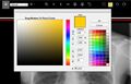 Figure 1.6 ODIN Colour Palette Toolbar.jpg 1,459 × 942; 154 KB
-
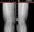 Figure 1.7a ODIN Angle Measurement Tool.jpg 1,426 × 1,328; 221 KB
-
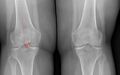 Figure 1.7b ODIN Angle Measurement Tool (closeup).jpg 1,850 × 1,150; 276 KB
-
 Figure 1.8 ODIN “unselect” annotation tool.jpg 1,036 × 1,056; 148 KB
-
 Figure 1.9 ODIN Download Image with Annotations.jpg 483 × 96; 4 KB
-
 Figure 10.10A Axial CT of Abdomen.jpg 1,272 × 744; 117 KB
-
 Figure 10.10B Coronal CT of Abdomen.jpg 762 × 794; 95 KB
-
 Figure 10.11A Abdominal x-ray of Toxic Megacolon.jpg 706 × 942; 88 KB
-
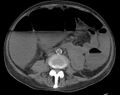 Figure 10.11B Axial CT of Toxic Megacolon.jpg 1,146 × 910; 109 KB
-
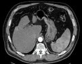 Figure 10.12A Axial CT of Liver.jpg 1,138 × 886; 151 KB
-
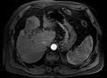 Figure 10.12B Axial MRI of Liver.jpg 1,298 × 950; 115 KB
-
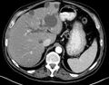 Figure 10.13A Axial CT of the liver demonstrating biliary system dilation.jpg 1,164 × 912; 168 KB
-
-
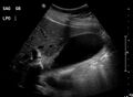 Figure 10.1A Sagittal ultrasound of the Gallbladder.jpg 1,474 × 1,076; 116 KB
-
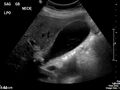 Figure 10.1B Sagittal Ultrasound of the Gallbladder Neck and Calculus.jpg 1,490 × 1,118; 132 KB
-
-
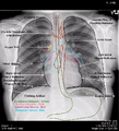
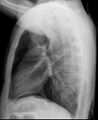 Figure 10.2B Lateral chest x-ray displaying sub-diaphragmatic free gas.jpg 1,004 × 1,228; 152 KB
-
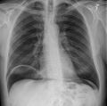 Figure 10.3A Chest x-ray displaying massive pneumoperitoneum.jpg 1,184 × 1,178; 180 KB
-
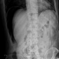 Figure 10.3B Decubitus Abdominal x-ray displaying massive pneumoperitoneum.jpg 1,202 × 1,198; 173 KB
-
-
-
-
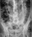
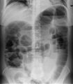
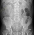 Figure 10.5B Abdominal x-ray, upright, suspicious for localized ileus.jpg 1,110 × 1,162; 162 KB
-
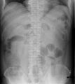
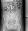 Figure 10.6A Abdominal x-ray, supine, suspicious for SBO.jpg 541 × 598; 181 KB
-
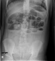 Figure 10.6B Abdominal x-ray, upright, suspicious for SBO.jpg 538 × 586; 175 KB
-
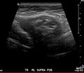 Figure 10.7A Ultrasound of Abdomen displaying a mass in the abdominal wall fat.jpg 1,438 × 1,244; 140 KB
-
-
-
-
-
-
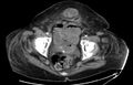
 Figure 11.1A Sagittal Ultrasound of the Uterus with a uterine mass.jpg 1,402 × 1,108; 139 KB
-
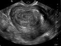 Figure 11.1B Transverse Ultrasound of the Uterus with a mass.jpg 1,446 × 1,092; 157 KB
-
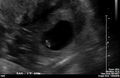
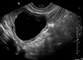 Figure 11.2A Pelvic ultrasound in a female with an ovarian cyst.jpg 1,506 × 1,092; 142 KB
-
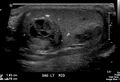
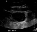 Figure 11.2B Sagittal ultrasound of a right ovarian cyst.jpg 1,338 × 1,170; 126 KB
-
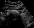 Figure 11.3A Ultrasound of female uterus, echogenic mass with acoustic shadowing.jpg 1,268 × 1,072; 136 KB
-
-
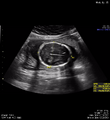
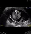
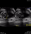
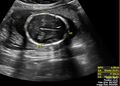 Figure 11.4A Fetal ultrasound demonstrating head circumference measurements.jpg 1,482 × 1,064; 178 KB
-
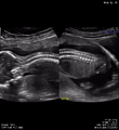
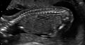 Figure 11.4B Fetal ultrasound demonstrating the spine.jpg 1,572 × 838; 132 KB
-

-
-
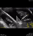
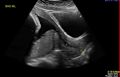 Figure 11.6A Sagittal midline ultrasound of the cervix and placenta.jpg 1,662 × 1,070; 128 KB
-
-
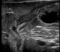
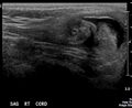
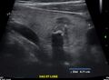 Figure 12.1A Sagittal Right Lobe Ultrasound of Thyroid.jpg 1,434 × 1,050; 144 KB
-
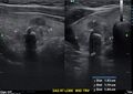 Figure 12.1B Sagittal Right Lobe Ultrasound of Thyroid.jpg 1,568 × 1,110; 200 KB
-
-
-
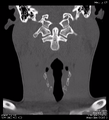
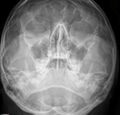
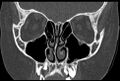
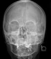
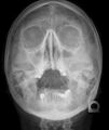
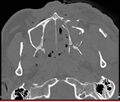 Figure 12.3A CT Scan of the Orbits and Facial region.jpg 1,274 × 1,086; 178 KB
-
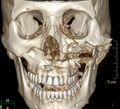 Figure 12.3B CT Scan 3D Rendering of Facial Bones.jpg 1,202 × 1,094; 206 KB
-
-
-
-
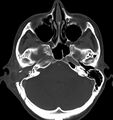
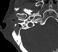 Figure 12.5B CT Scan of the Mastoid with air-cell opacity and skull erosion.jpg 1,300 × 1,174; 220 KB
-
-
-
-
 Figure 13.1 Ultrasound guided biopsy of a breast mass.jpg 1,342 × 1,032; 147 KB
-
 Figure 13.2 CT guided biopsy of a left lung mass.jpg 1,034 × 402; 57 KB
-
 Figure 13.3A CT scan of abdomen with a grid and measurements to plan drainage.jpg 1,498 × 1,046; 268 KB
-
 Figure 13.3B CT scan of a guided drainage of perihepatic abscess.jpg 1,046 × 750; 171 KB
-
-
-
 Figure 13.5 Chest x-ray of a PICC line.jpg 1,086 × 1,132; 151 KB
-
 Figure 13.7 Chest x-ray of a Tunneled Catheter.jpg 1,108 × 1,190; 154 KB
-
 Figure 13.8A Cordis IVC Filter.jpeg 372 × 640; 57 KB
-
 Figure 13.8B Cordis IVC Filter Placement in situ.jpg 932 × 1,182; 145 KB
-
-
-
-
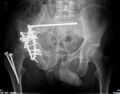 Figure 14.11B X-ray of the pelvis, post-operative fixation of pelvic fractures.jpg 1,434 × 1,124; 186 KB
-
-
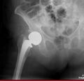 Figure 14.12B X-ray of the femur, post-operative arthroplasty.jpg 1,122 × 1,082; 142 KB
-
-
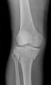
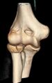
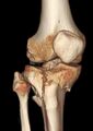 Figure 14.13B 3D image of the knee, displaying a complex tibial fracture.jpg 796 × 1,128; 79 KB
-
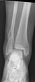 Figure 14.14A X-ray of the ankle, displaying fractures.jpg 418 × 960; 52 KB
-
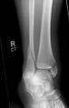 Figure 14.14B X-ray of the ankle, displaying fractures.jpg 630 × 990; 67 KB
-
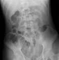
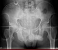
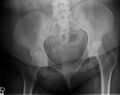 Figure 14.15A X-ray of the pelvis.jpg 823 × 653; 116 KB
-
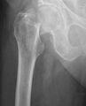
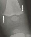
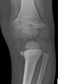
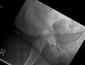 Figure 14.15B X-ray of the femur and hip joint.jpg 612 × 464; 55 KB
-
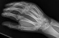
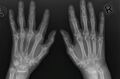 Figure 14.16A X-rays of both hands displaying erosive arthritis and joint deformity.jpg 1,534 × 1,014; 171 KB
-
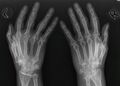 Figure 14.16B X-rays of both hands displaying erosive arthritis and joint deformity.jpg 1,410 × 1,010; 158 KB
-
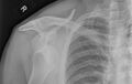
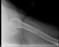
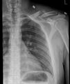 Figure 14.1A X-ray of the left shoulder, pre-operative, clavicle fracture.jpg 898 × 1,064; 123 KB
-
 Figure 14.1B X-ray of the left shoulder, post-operative, clavicle fracture.jpg 1,528 × 1,140; 144 KB
-

-
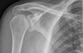
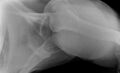
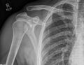 Figure 14.2B X-ray of the right shoulder, AP, with AC joint separation.jpg 1,390 × 1,074; 203 KB
-
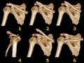 Figure 14.3 Acromioclavicular injury classification.jpg 1,024 × 768; 108 KB
-
-
-
-
-
-
-
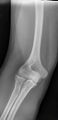 Figure 14.7A X-ray of the elbow displaying a radial head fracture.jpg 518 × 1,068; 61 KB
-
-
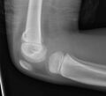
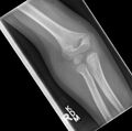 Figure 14.8A X-ray of the elbow displaying a humeral fracture.jpg 912 × 904; 83 KB
-
-
-
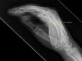
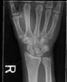
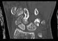 Figure 14.9B CT scan of the wrist displaying a scaphoid bone fracture.jpg 1,002 × 708; 75 KB
-
-
-
-

-
-
-
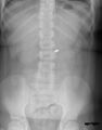 Figure 15.4A AP Abdominal x-ray, supine, of a foreign body.jpg 794 × 1,012; 101 KB
-
 Figure 15.4B Lateral abdominal x-ray, lateral, of a foreign body.jpg 710 × 972; 78 KB
-
 Figure 15.5A Inspiration chest x-ray for a suspected foreign body aspiration.jpg 1,104 × 1,008; 121 KB
-
-
-
-
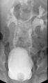
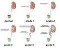 Figure 15.7 Vesico-ureteric reflux grading.jpg 990 × 803; 69 KB
-
-
-
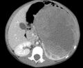 Figure 15.9B Axial CT of the abdomen revealing a very large mass.jpg 980 × 804; 92 KB
-
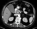
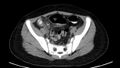
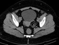
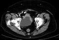
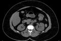 Figure 16.1A Axial CT of the abdomen.jpg 1,328 × 900; 144 KB
-
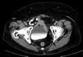
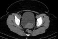 Figure 16.1B Axial CT of the pelvis displaying a left ureteric calculus.jpg 1,284 × 862; 141 KB
-
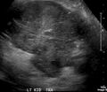 Figure 16.2A Transverse ultrasound of the left kidney mass.jpg 1,180 × 1,024; 132 KB
-
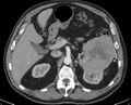 Figure 16.2B Axial CT of the abdomen revealing a renal tumour.jpg 1,260 × 1,020; 177 KB
-
-
-
-
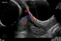
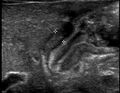
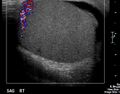
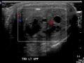 Figure 16.4B Transverse Doppler ultrasound of the left testicular tumour.jpg 1,326 × 1,000; 120 KB
-
-
-
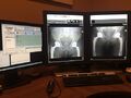 Figure 3.1 PACS Imaging Viewing Station (Workstation).jpg 3,264 × 2,448; 1.27 MB
-
 Figure 3.10 Common x-ray Test Object, Lucite Plastic Board.jpg 1,808 × 2,380; 994 KB
-
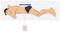 Figure 3.11 Decubitus positioning for a chest or abdomen x-ray.png 593 × 318; 23 KB
-
-
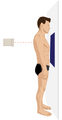
 Figure 3.11B Lateral, upright, chest x-ray positioning.png 426 × 730; 34 KB
-
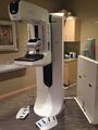 Figure 3.12 Mammography x-ray Machine.jpg 2,448 × 3,264; 1.34 MB
-
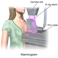 Figure 3.13A – CC Mammography Illustration.png 1,500 × 1,500; 1,011 KB
-
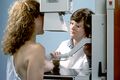 Figure 3.13B – Patient being positioned for a CC Mammogram.jpg 2,700 × 1,800; 2.38 MB
-
-
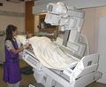 Figure 3.15 A Fluoroscopy Machine.jpg 398 × 324; 37 KB
-
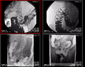 Figure 3.16 Barium Enema images.png 1,044 × 828; 746 KB
-
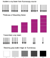 Figure 3.18 Fluoroscopy Images are Inverted in Comparison to x-rays.png 1,026 × 1,190; 273 KB
-
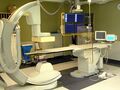 Figure 3.19 Angiography Machine with C-Arm.jpg 2,560 × 1,920; 872 KB
-
 Figure 3.2 – MRI Request Form.jpg 1,500 × 1,932; 1.72 MB
-
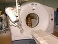 Figure 3.21 Helical CT Scanner.jpg 2,560 × 1,920; 679 KB
-
 Figure 3.25A Brain Level and Window.png 283 × 509; 50 KB
-
 Figure 3.25B Bone Level and Window.png 340 × 548; 62 KB
-
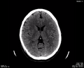 Figure 3.26A Head CT visible on brain level and window. Bone not well seen.png 1,002 × 815; 352 KB
-
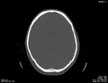 Figure 3.26B Head CT on bone level and window. Brain not well seen.png 1,002 × 766; 327 KB
-
 Figure 3.27 Standard viewing orientation for an axial CT image.png 1,002 × 766; 379 KB
-
 Figure 3.28A CT image displayed in axial orientation.png 1,002 × 815; 273 KB
-
 Figure 3.28B CT image displayed in sagittal orientation.png 1,002 × 766; 166 KB
-
 Figure 3.28C CT image displayed in coronal orientation.png 1,002 × 766; 182 KB
-
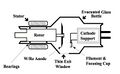 Figure 3.3 X-ray Tube, Schematic.jpg 721 × 445; 53 KB
-
-
-
 Figure 3.32A Ultrasound Machine.jpg 320 × 240; 20 KB
-
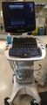 Figure 3.32B Mobile Ultrasound Machine.jpg 288 × 637; 55 KB
-
-
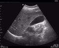 Figure 3.34 Gallbladder.png 1,002 × 815; 373 KB
-
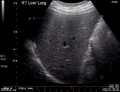 Figure 3.35A Normal Liver Ultrasound.png 1,002 × 766; 514 KB


.jpg)
.jpg)


.jpg)
.jpg)
.jpg)
.jpg)

.jpg)
.jpg)


.jpg)


.jpg)


.jpg)
.jpg)
.jpg)
.jpg)