File:1-s2.0-S0960896621006052-gr1.jpg
Jump to navigation
Jump to search

Size of this preview: 456 × 600 pixels. Other resolutions: 182 × 240 pixels | 527 × 693 pixels.
Original file (527 × 693 pixels, file size: 213 KB, MIME type: image/jpeg)
Summary
| Description |
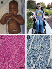
English: Fig. 1. X-linked myotubular myopathy. (A) 1 year old boy with genetically confirmed XLMTM. Note the long, myopathic facies, bilateral ptosis and extremity muscle hypoplasia. There is evidence of multiple technology dependencies, including invasive ventilator support and G tube for feeding. (B) 8 year old with genetically confirmed XLMTM and typical clinical features of facial weakness, extremity muscle hypoplasia, and wheelchair and ventilator requirements. (C, D) Representative photomicrographs from a muscle biopsy of a boy with confirmed XLMTM. (C) Hematoxylin and eosin stain, showing myofiber hypotrophy and many fibers with prominent, centrally located nuclei, (D) NADH stain showing numerous fibers with accumulated perinuclear organelles. Scale bar = 50 µm. |
| Date | |
| Source | https://www.sciencedirect.com/science/article/pii/S0960896621006052 |
| Author | Michael W. Lawlor , James J. Dowling |
Licensing
English: This file is licensed CC BY-NC-ND 4.0
This file was uploaded with UploadWizard.
File history
Click on a date/time to view the file as it appeared at that time.
| Date/Time | Thumbnail | Dimensions | User | Comment | |
|---|---|---|---|---|---|
| current | 22:29, 10 April 2023 | | 527 × 693 (213 KB) | Ozzie10aaaa (talk | contribs) | Uploaded a work by Michael W. Lawlor , James J. Dowling from https://www.sciencedirect.com/science/article/pii/S0960896621006052 with UploadWizard |
You cannot overwrite this file.
File usage
There are no pages that use this file.