File:PMC4898078 gr2.png
Jump to navigation
Jump to search
No higher resolution available.
PMC4898078_gr2.png (512 × 506 pixels, file size: 155 KB, MIME type: image/png)
Summary
| Description |
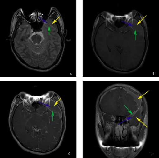
English: fig2: 22-year-old man with complicated arachnoid cyst. A, Axial T2-weighted MR image. The lesion is transected in the middle by the arachnoid membrane (blue arrow). Both segments (green and yellow arrows) demonstrate signal that is isointense to the adjacent brain parenchyma. B, Axial T1-weighted MR imaging better demonstrates the lesion’s complex nature. The lesion represents an arachnoid cyst (green arrow) which shows evidence of hemorrhage as demonstrated by the presence of T1 shortening. The surrounding subdural hematoma (yellow arrow) shows well demarcated prominent T1 shortening indicative of hemorrhage. C, Contrast enhanced axial T1-weighted MR imaging shows enhancement of the dural membrane but does not show enhancement within the arachnoid cyst (green arrow). The arachnoid membrane (blue arrow) separates hemorrhage in the subdural compartment (yellow arrow) from hemorrhage inside the arachnoid cyst (green arrow). D, Coronal contrast enhanced T1-weighted MR imaging shows the arachnoid cyst (green arrow), arachnoid membrane (blue arrow), and subdural hematoma (yellow arrows). |
| Date | |
| Source | https://openi.nlm.nih.gov/detailedresult?img=PMC4898078_gr2&query=&req=4 |
| Author | Patel AP, Oliverio PJ, Kurtom KH, Roberti F |
Licensing
English: This file is licensed CC BY-NC-ND 4.0
This file was uploaded with UploadWizard.
File history
Click on a date/time to view the file as it appeared at that time.
| Date/Time | Thumbnail | Dimensions | User | Comment | |
|---|---|---|---|---|---|
| current | 21:34, 19 February 2023 | | 512 × 506 (155 KB) | Ozzie10aaaa (talk | contribs) | Uploaded a work by Patel AP, Oliverio PJ, Kurtom KH, Roberti F from https://openi.nlm.nih.gov/detailedresult?img=PMC4898078_gr2&query=&req=4 with UploadWizard |
You cannot overwrite this file.
File usage
There are no pages that use this file.